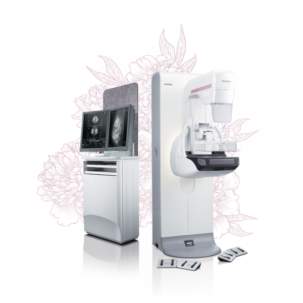
Innovation and quality in mammography
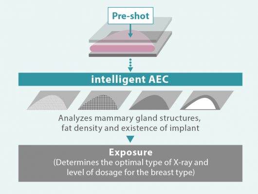
AMULET Innovality – the result of Fujifilm’s ongoing “innovation” and commitment to providing top “quality” mammography services. The Innovality utilises Fujifilm’s unique a-Se direct conversion flat panel detector (FPD)*1 to produce clear images with a low X-ray dose. This system makes use of intelligent AEC (i-AEC) combined with a image analysis technology to automatically adjust the X-ray dosage for each breast type. AMULET Innovality is a highly advanced mammography system which offers an extremely fast image interval of just 15 seconds. With this system, Fujifilm furthers the provision of high quality examinations with superior image quality.
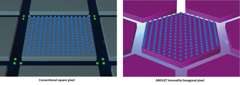
*1 Using a HCP (Hexagonal Close Pattern) TFT array.
Unique detector for fast, low dose examinations
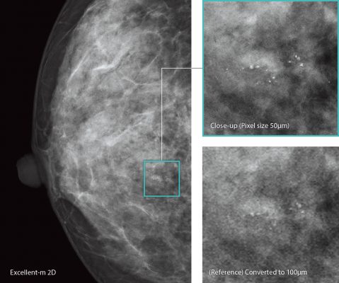
AMULET Innovality employs a direct-conversion flat panel detector made of Amorphous Selenium (a-Se) which exhibits excellent conversion efficiency in the mammographic X-ray spectrum. The HCP (Hexagonal Close Pattern) detector efficiently collects electrical signals converted from X-rays to realize both high resolution and low noise. This unique design makes it possible to realize a higher DQE (Detective Quantum Efficiency) than with the square pixel array of conventional TFT panels. With the information collected by the HCP detector, AMULET Innovality creates high definition images with a pixel size of 50 μm; the finest available with a direct-conversion detector.
This low-noise and high-speed switching technology allows tomosynthesis exposures with a low X-ray dosage and short acquisition time to be performed. Fast image display is also possible, realizing a smooth mammography workflow from exposure to image display.
ISC – Adjusted contrast and low X-ray dose using a Tungsten Target
Image-based Spectrum Conversion*1 (ISC) technology can be used to adjust contrast in an image. ISC analyzes images to compensate for variations in contrast due to the density of mammary glands, amount of fat and X-ray spectrum. ISC aims to ensure that images display adequate contrast even with the use of a high energy, low-dose X-ray beam. This technology allows sites that previously exploited the superior contrast of a Molybdenum target to realize the dose advantages offered by the use of Tungsten without having to compromise image contrast.
*1 Based on Image analysis the appearance is adjusted to emulate the image quality with the simulated “optimal” spectrum.
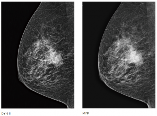
DYN II – Provides high contrast image without saturation in breast region
Dynamic Visualization II (DYN II) provides consistent appropriate density of glandular and adipose tissue in each breast type, so the contrast of thick breast and dense breast is improved. Furthermore, it provides high contrast with no saturation in breast region, so the sites are possible to set high contrast parameter.
Unique detector for fast, low dose examinations
intelligent AEC has advantages in defining the appropriate dose for an examination compared to conventional AEC systems where the sensor position is fixed. Through the analysis of information obtained from low- dose preshot images, intelligent AEC makes it possible to consider the mammary gland density (breast type) when defining the x-ray energy and level of dose required. Able to be used even in the presence of implants; intelligent AEC enables more accurate calculation of exposure parameters than is possible with conventional AEC systems. By allowing the use of automatic exposure for the implanted breast, intelligent AEC can further enhance examination workflow.
Related products
Digital Mammography

