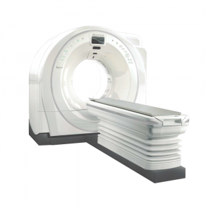
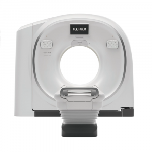
The new trend of CT Scanner
The needs for faster and more accurate diagnosis are increasing every days in the front line of medical practice.
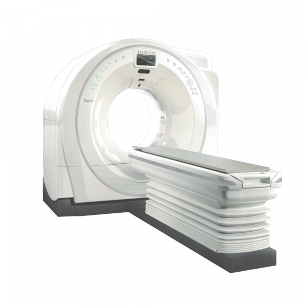
Supria is designed to answer in one CT all the demands for various routine applications, compact, useful results and ease of use without any compromise. Supria CT is your answer to take off to the next clinical and technology.
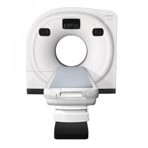
The top-notch gantry design with the compact body and the 75cm widest bore opening among the 16ch/32slices systems.
Iterative processing for routine examinations
To use the iterative processing (Intelli IP), a noise reduction technology, more efficiently on routine examinations, reconstruction speed has been improved by 50% compared to the Conventional CT. In addition, the intensity of noise reduction can be selected from seven levels, providing high quality images with appropriate exposure dose, image noise reduction and artifact reduction based on the operation
guideline at clinical sites.
High throughput, high image quality
High performance, such as high-speed rotation, submillimeter slice imaging, powerful X-ray generator and state-of-the-art image reconstruction algorithms,
realizes high resolution and high throughput examinations.

Submillimeter slice imaging for high resolution and high quality images
Supria realizes high resolution images in a short time based on 0.625mm x 16ch scanning. In addition, high resolution and smooth 3D images and MPR images can be achieved by submillimeter slice scanning. Oblique images by MPR can be also achieved after scanning.

HiMAR reduces metal artifacts
HiMAR (High Quality Metal Artifact Reduction) adopts unique algorithms for estimating and correcting artifacts based on metal data.

Capable of imaging in various patient’s positions
With a large bore of 750mm and a maximum effective field of view that reduces anxiety of the patient, it is possible to scan with various patient’s positions.

Helpful function to reduce the burden on the patient
Equipped with a motion artifact correction, body movement can be compensated even after scanning. Even if the patient is out of the effective field of view, such as a patient with a kyphosis, images can be reconstructed without re-scanning in case it is within the maximum effective field of view.

Motion artifact correction

Full FOV data acquisition

